Limb Lengthening Surgery

Limb Lengthening was first described in literature by Codvilla (1905) and Magnusion (1908). Since then many other surgeons including Ombredanne (1913), Putti (1921), Abbot (1927) performed limb lengthening using different techniques with varying success. Bost and Larsen (1930) were the first to performed femoral lengthening over an Intramedullary road after creation of osteotomy with a power or Gigli Saw.
In 1936, Anderson reported several innovations for femoral lengthening including the use of wires attached to the apparatus under tension and a technique for percutaneous osteotomy. From 1970 to 1990, Wagner method of lengthening became more popular than Anderson among most paediatric orthopaediats. This involved a three stage technique of rapid distraction of 2mm per day, bone grafting and plating with fixator removal.
However, it was Professor Ilizarov from remote Kugan, Siberia in the early 1950s who introduced the concept of local bone formation with a minimally invasive procedure, Ilizarov coined. The term distraction osteogenesis to describe the induction of new bone formation between osseous surfaces that are gradually pulled apart. He worked in a relatively isolated area of the world using distraction osteogenesis to treat a variety of musculoskeletal conditions.
The reconstruction of bone defects from post-trauma causing shortening and deformity was the broadest application of his method. Bone transport was used to salvage many limbs that otherwise would have been amputated because of non-union osteomyalitis or severe segmented bone loss. Later, Ilizarov used this method for limb lengthening in excess of 15 to 30 percent. He successfully lengthened several dwarfs.
During the lengthening procedure, the soft tissue including muscle nerves and blood vessels also grow in response to bone lengthening. The regenerate bone is normal and patients can walk or run eventually. His clinical success led to the spread of his work initially to the communist block of countries. By 1981, a group of Italian orthopaedic surgeons learned his technique and spread it to the West.
The technique arrived in Asia since the late 1980s. In some developing countries in Asia, the incidence of infected non-union and congenital deformities is higher resulting in vast experience in the Ilizarov technique. Advanced technique of limb lengthening and reconstruction for dwarfism, trauma or tumour replacement are also widely used in more developed countries in Asia.
The procedure can be performed both in children and adults with limb length discrepancy and deformity. In children limb shortening gives rise to limping, secondary scoliosis, increased energy expenditure and psychological problems. When the condition is not treated and continues to adulthood arthritis of the opposite knee, and hip and chronic back pain can develop.
In children, we use it commonly for conditions including proximal focal femoral deficiency, fibular hemimelia, congenital hemiatrophy. Growth plate defects or damage due to trauma or infection can cause shortening with deformity requiring treatment. In adults indication for limb lengthening include achondroplasia fibular, hemimelia, post-trauma shortening and poliomyelitis. Upper limb lengthening for humeral and thumb lengthening are occasionally performed.
Other common indications for Ilizarov technique include correction of deformities and non-union. This can be done acutely or with gradual correction. Bone transport is commonly used for treating non-union. With infected non-union, it is possible to resect large segments of infected bone and reconstruct the defect with bone transport.
The technique has also been used for reconstruction of feet with a variety of conditions including neglected club foot, complex foot deformities, equinus, ankle arthrodesis and metatarsal lengthening. The results are good and patient can walk with plastigrade foot.
The Ilizarov method is also an excellent method for treatment of acute unstable fractures with substantial bone loss. Intraarticular fractures of the tibia using minimally invasive external fixators and supplementary screw fixation have produced good results in terms off bony union and knee motion.
Of special motion is lengthening for constitutional short stature. These patients are a special group where careful psychological assessment is required prior to surgery. Guidelines for minimum height vary but generally are maximum height of 155 cm for females and 162 cm for males. Internal bone lengthening using expandable nails are preferred to minimize complications.
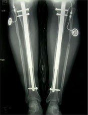
A variety of implants are available to achieve the desired goal including small wire external fixators, lengthening-over-nail techniques and internal expandable nails like Fitbone or ISKD.
Learn more about Limb Lengthening Surgery

Intramedullary Skeletal Kinetic Distractor (ISKD) is a technique in which the rod is inserted and screwed to the bone. It involves one direction to rotate the leg and the rod lengthens, expanding the leg and thus allowing the new bone to develop gradually.
 Learn more

Fitbone is an intelligent “state-of-the-art” nail where distraction is performed by an electronically controlled distractor set, including a power unit and a two-part guide nail. The operation of the system is regulated transcutaneously by a portable mini-radio control device.
 Learn more

uring the initial surgery, a metal rod is inserted into the central cavity (intramedullary) of the lower legs (the tibia bone), and then the external fixator device is attached to the bone. As the limb is lengthened, one end of the bone slides over the rod and new bone is grown around it.
 Learn more

A new technique, lengthening and then nailing (LATN), is effective in correcting leg length discrepancy (LLD) for a variety of conditions. The LATN technique uses monolateral frames for the distraction phase. Pins and wires are placed to allow subsequent intramedullary nailing.
 Learn more


 Adult Orthopaedic